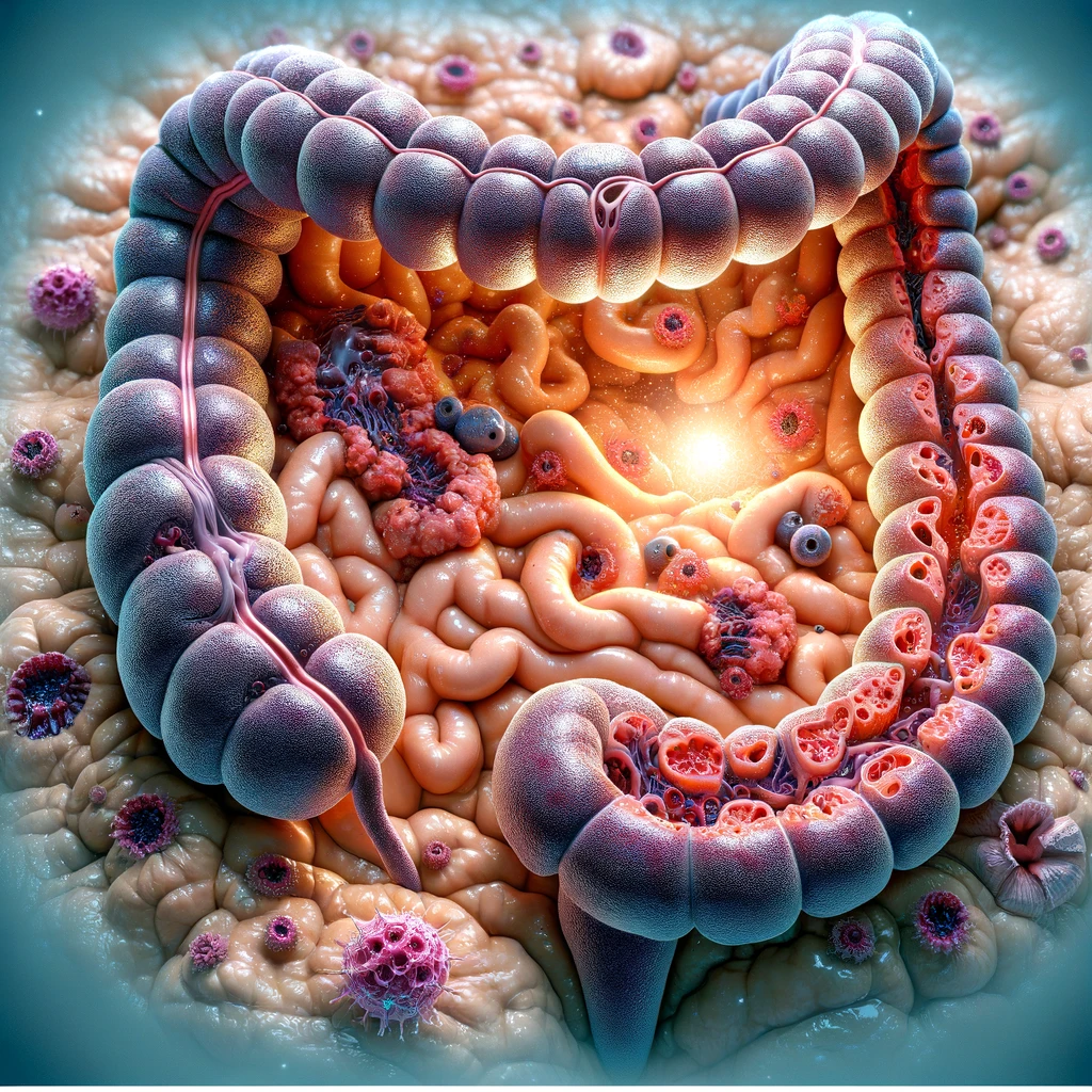
Gastrointestinal Stromal Tumor (GIST)

Gastrointestinal Stromal Tumor (GIST) is a rare type of tumor that occurs in the gastrointestinal tract, which includes the stomach, small intestine, and esophagus. It originates from the specialized cells called interstitial cells of Cajal (ICCs), also known as the pacemaker cells, that regulate the movement of food through the digestive system.

What is a GIST Tumor?

GISTs can be both benign (noncancerous) or malignant (cancerous). Malignant GISTs have the potential to spread to nearby tissues or metastasize to distant organs. These tumors are typically identified through imaging tests and confirmed by biopsy.

Common symptoms of GIST may include abdominal pain, blood in stool, anemia, difficulty swallowing, and unintended weight loss. However, some individuals with GIST may not experience any noticeable symptoms.

Treatment options for GIST mainly revolve around surgical removal of the tumor whenever possible. Additional therapies such as targeted drug therapy (tyrosine kinase inhibitors) may be used to control the growth and spread of malignant GISTs or when surgery is not feasible.

Beneficial Insights

Zovirax, Daklinza, and Addyi are all prescription drugs used in the treatment of specific medical conditions – Zovirax for herpes infections, Daklinza for hepatitis C, and Addyi for hypoactive sexual desire disorder in women.

It is essential to consult a healthcare professional for proper diagnosis, treatment, and ongoing management of Gastrointestinal Stromal Tumor.

Gastrointestinal Stromal Tumor (GIST)

  • Abdominal pain
  • Loss of appetite
  • Unexplained weight loss
  • Nausea and vomiting
  • Fatigue
  • Bloody stools or vomiting blood
  • Feeling of fullness after eating small amounts
  • Swelling or lumps in the abdomen
  • Anemia (low red blood cell count)
  • Difficulty swallowing or digesting food
  • Intestinal obstruction

Gastrointestinal Stromal Tumor (GIST)

  • Causes:
  • GIST is primarily caused by genetic mutations in the KIT or PDGFRA genes.
  • These mutations lead to the uncontrolled growth and division of cells in the gastrointestinal (GI) tract.
  • While the exact cause of these genetic mutations is unclear, they may occur spontaneously or be inherited.
  • Exposure to certain chemicals or substances, such as high levels of radiation or certain medications, may also increase the risk of developing GIST.
  • However, the majority of GIST cases do not have a specific known cause.

Diagnosis Methods:

1. Physical Examination: A thorough examination of the abdomen is conducted by a healthcare professional to look for any abnormal findings, such as mass or tumor.

Physical Examination

A healthcare professional will conduct a thorough examination of the abdomen to check for any physical abnormalities or signs of a tumor.

2. Blood Tests: Certain blood tests, such as complete blood count (CBC) and liver function tests (LFTs), may be conducted to assess overall health and detect any abnormalities that could be associated with GIST.

Blood Tests

Blood samples will be taken and analyzed for a complete blood count (CBC) and liver function tests (LFTs) to evaluate overall health and identify any abnormalities that might be associated with GIST.

3. Imaging Tests:
a. Ultrasound: Sound waves are used to create images of the abdomen, helping to detect any tumors or abnormal growths.

Ultrasound

Sound waves are used to create images of the abdomen and assess the presence of tumors or abnormal growths.

b. CT Scan (Computed Tomography): A series of X-ray images taken from different angles are combined to create detailed cross-sectional images. This helps visualize the extent of the tumor, its size, and location.

CT Scan (Computed Tomography)

A series of X-ray images taken from different angles are combined to create detailed cross-sectional images of the abdomen, providing information about the size, location, and extent of the tumor.

c. MRI (Magnetic Resonance Imaging): Powerful magnets and radio waves are used to create detailed images of the abdomen. This can help in evaluating the extent and characteristics of the tumor.

MRI (Magnetic Resonance Imaging)

Powerful magnets and radio waves are used to create detailed images of the abdomen, which aid in evaluating the characteristics and extent of the tumor.

4. Endoscopy: A long, flexible tube with a camera attached (endoscope) is inserted into the patient's gastrointestinal tract through the mouth or rectum. This allows direct visualization of the tumor and collection of tissue samples for biopsy.

Endoscopy

A long, flexible tube with a camera attached (endoscope) is inserted into the gastrointestinal tract through the mouth or rectum, enabling direct visualization of the tumor and collection of tissue samples for biopsy.

5. Biopsy: Tissue samples are obtained from the suspected tumor site and sent to a laboratory for examination under a microscope, confirming the presence of GIST.

Biopsy

Tissue samples are collected from the suspected tumor site and sent to a laboratory for microscopic examination to confirm the presence of GIST.